ACL Repair NYC — Primary ACL Repair vs. Reconstruction at New York Bone & Joint
Written by: Dr. Leon Popovitz
Board-Certified Orthopedic Surgeon (ABOS) │ FAAOS │ Fellowship-Trained Sports Medicine, NYU Langone
Knee & Shoulder Surgery │ Co-Founder, New York Bone & Joint │ Surgery at Lenox Hill Hospital
Date Published: June 30, 2026
Last Updated: July 16, 2026
Last Medically Reviewed: July 16, 2026
This page has been written and reviewed by a fellowship-trained, board-certified orthopedic surgeon with over 20 years of clinical experience. All clinical claims are supported by peer-reviewed literature and current AAOS and AOSSM guidelines. See References section below.
At a glance
What it is: ACL primary repair is a surgical procedure where the torn native ACL is structurally sutured back together and reinforced with a bioactive scaffold (the BEAR® implant) to support the biological healing of your own tissue, rather than replacing it with a tendon graft.
Who performs it: Dr. Leon E. Popovitz, board-certified orthopedic surgeon (ABOS), FAAOS, fellowship-trained sports medicine NYU Langone. Surgery at Lenox Hill Hospital, outpatient.
Strict Candidacy Criteria: Proximal (femoral-side) tear location, acute timeline (ideally within 3 weeks of injury), excellent native tissue volume remaining, and no diffuse joint wear or extensive cartilage loss.
Who does NOT qualify: Mid-substance or distal tears. Chronic tears (> 3–4 weeks). Patients who have failed prior ACL surgery. Significant bone loss or concurrent structural damage. Most ACL tears fall outside the repair criteria. ACL reconstruction remains the standard of care for most patients. [4]
The BEAR® Implant: An FDA-approved bioactive bovine-collagen scaffold that is soaked in the patient’s own blood intraoperatively. It functions as a fluid-resistant bridge between the torn ends, retaining specialized growth factors to stimulate native ligament healing.
Clinical Track Record: Clinical Evidence: The landmark BEAR Trial (New England Journal of Medicine) demonstrated statistical non-inferiority to traditional autograft reconstructions at a 2-year clinical follow-up in highly selected cohorts. [1]
Access: Same-week consultations. No referral required. Both Manhattan locations.
Book a consultation
Prompt Appointments Available
Upper East Side: 1198 Third Ave | Midtown: 425 Madison Ave
Surgery performed at Lenox Hill Hospital - Northwell Health
About this page
Written by Dr. Leon Popovitz, board-certified orthopedic surgeon (ABOS), FAAOS, fellowship-trained sports medicine NYU Langone. Co-founder New York Bone & Joint Specialists, Manhattan, New York City. ACL repair procedures performed at Lenox Hill Hospital — Northwell Health, as outpatient surgery. This page addresses ACL primary repair specifically. For ACL reconstruction — the standard of care for most ACL tears — see acl-reconstruction-nyc.
Why the ACL Cannot Repair Itself
The anterior cruciate ligament (ACL) possesses a specific, highly frustrating biological limitation that sets it apart from almost every other ligament in the musculoskeletal system.
When an extra-articular ligament like the Medial Collateral Ligament (MCL) tears, it forms a natural blood clot that acts as a scaffold, allowing the tissue to heal and bridge reliably on its own. The ACL behaves completely differently due to its intra-articular environment. [2]
Retraction and Synovial Bathing: Upon tearing, the ends of the ACL immediately snap back and retract. They are then continuously bathed in synovial fluid, the lubricating fluid that fills the inner knee joint cavity.
Dissolution of the Fibrin Scaffolding: Synovial fluid contains specialized enzymes that rapidly dissolve the delicate fibrin blood clots that the body naturally attempts to produce following an injury.
Failure to Bridge: Without an uninterrupted biological scaffold, the torn ends of the ACL are physically incapable of bridging the gap to mend themselves.
Traditional ACL surgery bypasses this issue entirely by executing an arthroscopic ACL reconstruction to completely replace the native tissue with a new graft. A primary ACL repair, by contrast, attempts to neutralize this fluid barrier by introducing the dense BEAR® implant. This acts as a physical shield and bioactive bridge that resists synovial degradation long enough for your own cells to safely cross and repair the native ligament. [3]
ACL Primary Repair vs. ACL Reconstruction: Side by Side
| ACL Primary Repair (BEAR®) | ACL Reconstruction | |
| Surgical Mechanism | Suture repair of native tissue reinforced with a bioactive collagen scaffold. | Complete excision of the torn ligament, replaced with a tendon autograft or allograft. |
| Native Tissue Preservation | High; the native ligament is fully preserved and retains its natural nerve receptors. | Native ACL is completely removed and replaced by a substitute tendon graft. |
| Candidacy Envelope | Exceptionally narrow: acute proximal tears, within 3 weeks of injury, rich tissue volume. [5] | Broad; appropriate for nearly all complete ACL tears in active individuals. |
| Evidence | Promising short-term data; proven non-inferior at 2-year endpoints in selected cohorts. [1] | Decades of global clinical tracking. Widely considered the absolute gold standard. |
| Recovery | Approximately 9 months of criteria-based sports physical therapy. | Approximately 9 months of progression-based rehabilitation. |
| Re-tear risk | Eliminates graft harvest discomfort; preserves natural joint proprioception. | Highly predictable, structurally stable, and versatile across all tear patterns. |
| Best suited for | Re-tear Risk | Best Suited For |
References
- Murray MM et al. Bridge-Enhanced Anterior Cruciate Ligament Repair Is Not Inferior to Autograft Anterior Cruciate Ligament Reconstruction at 2 Years. Am J Sports Med. 2020. pubmed.ncbi.nlm.nih.gov/32298131
- Murray MM et al. Enhanced histologic repair in a central wound in the anterior cruciate ligament with a collagen-platelet-rich plasma scaffold. J Orthop Res. 2007. pubmed.ncbi.nlm.nih.gov/17415785
- Murray MM et al. The Bridge-Enhanced Anterior Cruciate Ligament Repair (BEAR) Procedure. Orthop J Sports Med. 2016. pubmed.ncbi.nlm.nih.gov/27900338
- AAOS. ACL Injury. OrthoInfo. orthoinfo.aaos.org/en/diseases–conditions/anterior-cruciate-ligament-acl-injuries
- Kon E et al. Biologic agents to optimize outcomes following ACL repair and reconstruction. J Orthop Res. 2022. pubmed.ncbi.nlm.nih.gov/33586785
FAQs
Yes, but only within a highly precise, narrow window of carefully selected candidates presenting with acute proximal ACL tears. [5] Primary repair utilizing the BEAR® (Bridge-Enhanced ACL Restoration) technique represents a sophisticated, emerging track that delivers excellent results for the correct anatomy. For the vast majority of presentation patterns (including mid-substance tears, chronic tissue scarring, or revision settings) undergoing an arthroscopic ACL reconstruction remains the undisputed standard of care. [4]
The BEAR® procedure involves using an FDA-approved bioactive collagen matrix scaffold that is saturated with the patient’s own fresh blood inside the operating suite. [3] The construct is carefully anchored between the torn native ACL stumps during a baseline knee arthroscopy session. This scaffold functions as a chemical shield that bridges the structural gap, while the concentrated blood delivers vital growth factors that stimulate the native ligament to heal itself.
The eligibility matrix is strict: the tear must be localized specifically at or near the femoral bone attachment (proximal), the injury must be acute (ideally evaluated and operated on within 3 weeks of the initial tear), there must be a clean, high-volume tissue stump remaining to physically accept sutures, and the joint must be free from extensive, concurrent structural damage. [5] Dr. Popovitz reviews your high-resolution MRI and movement parameters to determine if these criteria are met.
Current clinical evidence does not support the claim that a repair is broadly superior to a standard reconstruction. The landmark BEAR Trial proved statistical non-inferiority at two years, which indicates that it provides comparable functional outcomes, not superior ones. [1] The correct clinical perspective is that for a highly specific patient with a fresh, proximal tear, a primary repair offers an excellent, graft-sparing alternative that successfully preserves your native tissue and nerve pathways.
If a primary ACL repair fails to fully heal or tears again down the road, it does not preclude you from having future procedures. [4] Because the native anatomy and bone pathways are safely preserved during the repair attempt, a standard reconstructive ACL surgery using an autograft or allograft can be smoothly executed as a secondary procedure. This provides significant peace of mind to patients weighing their initial tracking choices.
The rehabilitation timelines are remarkably similar: both require roughly 9 months of protected, milestone-driven physical therapy before returning to high-impact cutting or pivoting sports. [1] The biological time required for a repaired native ligament to securely knit back together is nearly identical to the timeline required for a tendon graft to undergo full ligamentization inside the joint. Your milestones will be validated through strict objective functional testing rather than a calendar date.
Yes. If an acute sports injury causes a complex combination of damage, a primary repair can be performed concurrently alongside an arthroscopic meniscus repair or excision surgery or a localized cartilage preservation step if needed. [5] However, if the knee presents with extensive, multi-ligament instability or diffuse damage, a standard reconstruction track is almost always favored to ensure total mechanical stability.
Medically Reviewed by Dr. Popovitz.
You can book your appointment with New York Bone & Joint online by submitting the appointment request form here:
Book an appointmentOur Locations
-
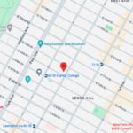
Upper East Side: Full Service Orthopedic Center & Walk-In
1198 3rd Avenue, Between 69th and 70th Street New York, NY 10021Physician appointments: Mon–Fri 9am–5:30pm, Sat 8:30am–4:30pm
Physical therapy: Mon–Fri 7am–7pm, Sat 8am–2pmPhone: (212) 759-4553 -

Orthopedic Doctor Midtown Manhattan NYC — New York Bone & Joint Specialists
425 Madison Ave, Suite 200 (second floor) New York, NY 10017 (corner of East 49th Street)Physician appointments: Mon–Fri 8:00 am–6:00 pm
Physical therapy: Mon–Fri 7 am–7 pm, Sat 8 am–2 pmPhone: (212) 759-4553 -
Orthopedic Doctor Upper East Side NYC — New York Bone & Joint Specialists
130 E 67th St New York, NY 10065Physician appointments: Mon–Fri 8:00am–6pm, Sat 8:30am–4:30pm
Physical therapy: Mon–Fri 7am–7pm, Sat 8am–2pmPhone: (212) 759-4553