Cartilage Restoration NYC — Knee Cartilage Repair at New York Bone & Joint
Written by: Dr. Popovitz.
Board-Certified Orthopedic Surgeon | Fellowship-Trained Sports Medicine, NYU Langone Medical Center | FAAOS
Co-Founder, NY Bone & Joint Specialists | Lenox Hill Hospital - Northwell Health
Recognized: New York Magazine Best Doctor | New York Times Super Doctor | IAOS Leading Physician of the World | IAOS Top Orthopedic Surgeon in New York
Date Published: June 29, 2026
Last Updated: June 29, 2026
Last Medically Reviewed: June 29, 2026
This page has been written and reviewed by a fellowship-trained, board-certified orthopedic surgeon with over 20 years of clinical experience. All clinical claims are supported by peer-reviewed literature and current AAOS and AOSSM guidelines. See References section below.
At a glance
What it is: Cartilage restoration is a group of surgical procedures designed to repair or replace damaged articular cartilage in the knee, preserving the patient’s own joint by regenerating or transplanting tissue. [1]
Who performs it: Dr. Leon E. Popovitz, board-certified orthopedic surgeon (ABOS), FAAOS, fellowship-trained sports medicine NYU Langone. Surgery at Lenox Hill Hospital, outpatient.
Procedures available: Microfracture (<2 cm² defects), OATS/mosaicplasty (1–3 cm² autografts), OCA (>3 cm² allografts), and MACI (2–10 cm² laboratory cell cultures). [1]
Best candidates: Young to middle-aged active patients (typically under 50) with focal, contained cartilage defects from injury rather than diffuse degenerative arthritis. Patients who have been told they are ‘too young for knee replacement.’
Outcomes: OATS: 92% good-to-excellent results in appropriately selected patients. [3] MACI significantly superior to microfracture at 2 years for defects >3 cm². [5] OCA durable at 5–10 years for large defects in young active patients. [4]
Recovery: Microfracture: 6–9 months to sport. OATS: 6–12 months. MACI: 12–18 months. All criteria-based clearance.
Access: Same-week consultations. No referral required. Both Manhattan locations.
Book a consultation
Prompt Appointments Available
Upper East Side: 1198 Third Ave | Midtown: 425 Madison Ave
Surgery performed at Lenox Hill Hospital - Northwell Health
About this page
About This Page: Written by Leon E. Popovitz, MD, board-certified orthopedic surgeon (ABOS), FAAOS, fellowship-trained sports medicine NYU Langone. Co-founder NY Bone & Joint Specialists, Manhattan, New York City. All cartilage restoration procedures at Lenox Hill Hospital — Northwell Health, as outpatient surgery.
Introduction
Articular cartilage is the smooth hyaline cartilage that coats the ends of the femur and tibia inside the knee joint, allowing the joint surfaces to glide against each other with minimal friction. It’s also one of the most biologically limited tissues in the body, possessing zero blood supply and zero capacity to regenerate spontaneously. [1]
When articular cartilage is damaged by acute trauma, repetitive loading, or a prior injury, the damage is permanent without surgical intervention. The cartilage surface does not heal, the bone beneath it is progressively exposed, and the joint space narrows. Over time, this progression leads to localized osteoarthritis. Cartilage restoration procedures interrupt this process by repairing the focal defect before the surrounding joint architecture deteriorates beyond repair.
The right procedure depends on defect size, location, and the underlying bone condition. The outcome depends on matching the procedure to the defect precisely.
Cartilage Damage: Understanding the Grade
| Grade | Description | Clinical Significance |
| Grade 1 | Surface softening and mild fibrillation. Intact surface structure. | Typically asymptomatic. No surgical intervention needed. |
| Grade 2 | Partial-thickness loss with fissuring <50% of the cartilage depth. | May produce symptoms. Non-surgical management usually appropriate. |
| Grade 3 | Deep fissuring >50% depth down to, but not through, the subchondral bone. | Clinically symptomatic. Cartilage restoration appropriate for focal contained defects. |
| Grade 4 (bone on bone) | Full-thickness cartilage loss entirely exposing the subchondral bone. | Highly symptomatic. Restorable if localized in young patients. Diffuse wear requires joint replacement options. |
Cartilage Restoration Procedures at New York Bone & Joint
Microfracture
Microfracture is the least invasive option.
Small holes are made in the subchondral bone beneath the defect to release bone marrow elements (such as stem cells and growth factors) that form a blood clot to fill the gap. [2]
The Biological Catch: This process forms fibrocartilage, which is rich in Type I collagen. Fibrocartilage is structurally softer, less durable, and less resistant to compressive loads than native hyaline cartilage, which is built from a dense matrix of Type II collagen optimized to withstand shear forces. Fibrocartilage acts more like a biological patch than a permanent shock absorber.
Candidacy: Clinical outcomes show excellent initial results that tend to decline at the five-year mark for larger lesions. [2] Microfracture is therefore reserved for small, contained defects (<2 cm²) in lower-demand scenarios.
OATS: Osteochondral Autograft Transfer
The OATS procedure involves harvesting a healthy cylindrical plug of bone and overlying cartilage from a non-weight-bearing peripheral zone of the patient’s own knee and transplanting it directly into the defect area. [3]
The Advantage: This technique transplants genuine, durable hyaline cartilage (rich in Type II collagen) into the injury zone rather than a fibrocartilage scar patch, providing a structurally superior repair material.
Candidacy: Published data demonstrates a 92% good-to-excellent success rate at intermediate follow-up. It’s ideal for focal defects measuring 1–3 cm² where a minimal number of autograft plugs can completely cover the area. Multiple plugs can be arranged in a mosaic pattern (mosaicplasty) for compound shapes. [3]
OCA: Osteochondral Allograft
Osteochondral allograft transplantation utilizes a precisely matched donor bone and cartilage plug obtained from a certified tissue bank, eliminating the need to harvest healthy tissue from the patient’s own knee. [4]
The Advantage: It completely eliminates donor-site harvest discomfort and allows for the treatment of large defects (>3 cm²) that require a larger volume of tissue than an autograft could safely provide.
Candidacy: The precisely measured graft is press-fit tightly into the defect. Intermediate studies show high graft durability at 5 to 10 years in highly active populations. [4]
MACI: Matrix-Induced Autologous Chondrocyte Implantation
MACI is an advanced, cell-based two-stage procedure. In the first stage, a minor biopsy of healthy cartilage is harvested arthroscopically. These native chondrocytes are isolated and multiplied in a specialized laboratory over a 4–6 week period. In the second stage, the expanded cellular matrix is seeded onto a structural collagen membrane and secured directly over the prepared defect area. [5]
The Advantage: In randomized controlled trials, MACI has shown statistically superior clinical outcomes compared to microfracture at two years for defects exceeding 3 cm², providing long-lasting hyaline-like tissue repair for large-scale, high-impact defects. [5]
Comprehensive Pre-Operative Evaluation: Protecting the Graft
Cartilage graft success depends on the mechanical environment surrounding it. Before any cartilage restoration procedure, Dr. Popovitz evaluates the entire mechanical alignment of the leg and the status of surrounding structures. If a malalignment such as a varus (bowlegged) or valgus (knock-kneed) deformity is present, a concurrent distal femoral or proximal tibial osteotomy may be planned to redistribute forces and protect the new graft.
Similarly, meniscal deficiency must be addressed simultaneously: a knee without a functioning meniscus transfers excessive compressive forces directly onto the cartilage graft, significantly increasing failure risk. When structural damage is caused by a sports injury, cartilage restoration may be performed concurrently with a meniscus repair or a stabilizing ACL reconstruction to ensure long-term joint health and protect the investment of the cartilage surgery.
Choosing the Right Procedure
| Procedure | Defect Size | Best Candidate | Recovery to Sport |
| Microfracture | < 2 cm² | Smaller focal defects. Older or lower-demand patients. Patients not candidates for more complex procedures. | 6–9 months |
| OATS | 1–3 cm² | Focal contained defects. Young active patients. Single or limited plugs can cover defect. | 6–12 months |
| OCA | > 3 cm² | Large focal defects. Young active patients. When autograft volume is insufficient. | 9–12+ months |
| MACI | 2–10 cm² | Larger focal defects in young, highly motivated patients. Patients who can commit to 12–18 month recovery. | 12–18 months |
References
- AAOS. Articular Cartilage Restoration. OrthoInfo. orthoinfo.aaos.org/en/treatment/articular-cartilage-restoration
- Becher C et al. The microfracture technique for the treatment of articular cartilage lesions. Orthopade. 2008. pubmed.ncbi.nlm.nih.gov/18288472
- Hangody L et al. Mosaicplasty for the treatment of articular cartilage defects. Clin Orthop Relat Res. 2001. pubmed.ncbi.nlm.nih.gov/11603716
- Matthews JR et al. Comparison of clinical outcomes following osteochondral allograft transplantation for osteochondral versus chondral defects in the knee. Knee Surg Relat Res. 2022. pubmed.ncbi.nlm.nih.gov/35509057
- Brittberg M et al. Treatment of deep cartilage defects with autologous chondrocyte transplantation. N Engl J Med. 1994. pubmed.ncbi.nlm.nih.gov/8078550
- Farr J et al. Clinical cartilage restoration: evolution and overview. Clin Orthop Relat Res. 2011. pubmed.ncbi.nlm.nih.gov/21240578
FAQs
Cartilage restoration is a group of specialized surgical techniques that repair, resurface, or replace localized zones of damaged articular cartilage inside the knee to halt degenerative breakdown and delay or completely avoid a premature joint replacement. [1] It is specifically designed for young to middle-aged active patients (typically under 50) with clear, localized cartilage defects resulting from an injury rather than extensive, widespread osteoarthritis.
No, articular cartilage is one of the few tissues in the human body with essentially zero capacity to heal or regenerate spontaneously because it does not possess a cellular blood supply. [1] Once the surface matrix is torn or worn away, it will not repair itself over time. Surgical restoration is required to structurally seal the surface using marrow stimulation, autograft transplants, allograft tissue, or laboratory-grown cellular implantation.
Microfracture punctures the bone to stimulate a fibrocartilage scar tissue patch to fill the gap, whereas the OATS procedure transplants structural plugs of genuine native hyaline cartilage and bone harvested from a low-stress area of your own joint. [2] [3] Hyaline cartilage is the optimal joint material you were born with; it is significantly harder, more resilient, and better adapted to bear weight than a softer fibrocartilage clot. The key difference is that hyaline cartilage is rich in Type II collagen optimized for compressive load, while fibrocartilage is predominantly Type I collagen, acting more like a biological patch.
MACI (Matrix-Induced Autologous Chondrocyte Implantation) is a multi-step biologic procedure where your own cartilage cells are harvested, multiplied across several weeks in a specialized lab environment, and then re-implanted into your knee defect via a protective collagen scaffold membrane. [5] MACI generates a robust, hyaline-like cartilage repair tissue that has shown statistically superior clinical outcomes versus traditional microfracture repairs for defects larger than 3 cm² in randomized controlled trials.
Timeline recovery varies based on the exact procedure performed: microfracture generally takes 6–9 months to return to sports; OATS takes 6–12 months; OCA takes 9–12+ months; and MACI requires an extended 12–18 month pathway for biological cell maturation. [1] Most protocols require partial or non-weight-bearing parameters for the initial 6–12 weeks to safely protect the healing graft while it integrates with the surrounding bone. Final athletic clearances are strictly criteria-based, utilizing objective functional testing and strength symmetry metrics rather than a standard calendar deadline.
Cartilage restoration was specifically developed as a treatment track for individuals in this exact predicament. If your knee has a clear, localized defect surrounded by otherwise healthy tissue, you don’t need an artificial metal joint. Dr. Popovitz performs an in-depth review of your mechanical axis, symptoms, high-resolution MRI, and surrounding structural health (including meniscal integrity and limb alignment) to map out an anatomical joint preservation surgery that restores your mobility safely. [1]
Yes, performing cartilage restoration concurrently with an ACL reconstruction or a meniscus surgery is highly common and clinically advantageous. [6] The marrow signals, stem cells, and localized growth factors released during the mechanical drilling of the ACL bone tunnels create an enriched biological healing environment that directly supports the integration of your cartilage graft, while allowing you to rehab both injuries along a synchronized timeline.
If an initial restoration option fails (such as a microfracture site breaking down or degrading over a five-year period), patients still have viable, joint-preserving options depending on the current stability of the bone. [2] Dr. Popovitz can perform a revision surgery using an escalating technique (such as moving from a failed microfracture to a structural OATS or MACI graft), introduce a mechanical alignment adjustment via a realigning osteotomy, or discuss localized partial replacement options if the wear has spread too far across the joint plane.
Medically Reviewed by Dr. Popovitz.
You can book your appointment with New York Bone & Joint online by submitting the appointment request form here:
Book an appointmentOur Locations
-
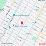
Upper East Side: Full Service Orthopedic Center & Walk-In
1198 3rd Avenue, Between 69th and 70th Street New York, NY 10021Physicians: Monday–Friday 8:00am–6:00pm, Saturday 8:00am–5:00pm
Physical Therapy: Monday–Friday 7:00am–7:30pm, Saturday 8:00am–3:00pm
Sunday: ClosedPhone: (212) 759-4553 -

Orthopedic Doctor Midtown Manhattan NYC — New York Bone & Joint Specialists
425 Madison Ave, Suite 200 (second floor) New York, NY 10017 (corner of East 49th Street)Physician appointments: Mon–Fri 8:00 am–6:00 pm
Physical therapy: Mon–Fri 7 am–7:30 pm, Sat 8 am–3 pmPhone: (212) 759-4553 -

Orthopedic Doctor Upper East Side NYC — New York Bone & Joint Specialists
130 E 67th St New York, NY 10065Tuesday & Thursday: 8:00am–6:00pm
All other days: ClosedPhone: (212) 759-4553