Patella Stabilization NYC — MPFL Reconstruction & Patellar Instability Treatment
Written by: Dr. Leon Popovitz
Board-Certified Orthopedic Surgeon (ABOS) │ FAAOS │ Fellowship-Trained Sports Medicine, NYU Langone
Knee & Shoulder Surgery │ Co-Founder, New York Bone & Joint │ Surgery at Lenox Hill Hospital
Date Published: June 30, 2026
Last Updated: July 13, 2026
Last Medically Reviewed: July 13, 2026
This page has been written and reviewed by a fellowship-trained, board-certified orthopedic surgeon with over 20 years of clinical experience. All clinical claims are supported by peer-reviewed literature and current AAOS and AOSSM guidelines. See References section below.
At a glance
What it is: Patella stabilization surgery repairs or reconstructs the stabilizing passive soft tissue restraints holding the kneecap in its femoral groove to halt recurrent lateral dislocations.
Who performs it: Dr. Leon E. Popovitz, board-certified orthopedic surgeon (ABOS), FAAOS, fellowship-trained sports medicine NYU Langone. Surgery at Lenox Hill Hospital, outpatient.
Procedures: MPFL Reconstruction (primary soft tissue restraint rebuild), Tibial Tubercle Osteotomy (TTT) (bony alignment shifting for high TT-TG distances), and Lateral Release (used strictly as an anatomical adjunct).
Who needs it: Patients with recurrent patellar dislocation (typically 2 or more episodes), young athletes after a first dislocation with high recurrence risk factors, and patients with chronic patellar instability limiting activity.
Recurrence rate without surgery: 30% to 50% re-dislocation rate after a first-time patellar dislocation if managed without surgical intervention. [2] This risk climbs with each successive episode.
Recovery: MPFL Reconstruction: 4–6 months to full sports return. Tibial Tubercle Osteotomy: 6–9 months to allow complete bony healing across the osteotomy site.
Access: Same-week consultations. No referral required. Both Manhattan locations.
Book a consultation
Prompt Appointments Available
Upper East Side: 1198 Third Ave | Midtown: 425 Madison Ave
Surgery performed at Lenox Hill Hospital - Northwell Health
About this page
Written by Dr. Leon Popovitz, board-certified orthopedic surgeon (ABOS), FAAOS, fellowship-trained sports medicine NYU Langone. Co-founder New York Bone & Joint Specialists, Manhattan, New York City. All patella stabilization procedures performed at Lenox Hill Hospital — Northwell Health, outpatient.
Introduction
A structural patellar dislocation occurs when the kneecap is forced entirely out of its anatomical groove on the femur, almost always displacing to the lateral side of the knee joint. The medial patellofemoral ligament (MPFL) serves as the primary passive mechanical restraint that checkmates this lateral displacement. Consequently, the MPFL is torn or severely attenuated in virtually every first-time traumatic patellar dislocation event. Once structurally compromised, the MPFL cannot reliably heal back to its native functional length or tension. This chronic laxity explains why the re-dislocation rate among young active patients approaches 30% to 50% without surgical intervention. [2]
Modern management of patellar instability requires a precise evaluation to identify the specific anatomical factors driving the joint failure. While most patients present with MPFL insufficiency as the primary driver, others possess structural bony anomalies—such as severe trochlear dysplasia (an abnormally shallow femoral groove that fails to contain the kneecap) or an elevated tibial tubercle offset—that must be addressed concurrently to prevent graft failure.
Surgical Patella Stabilization Tracks at New York Bone & Joint
MPFL Reconstruction
Medial patellofemoral ligament (MPFL) reconstruction biologically rebuilds the primary soft tissue checkrein on the inside of the knee. This is typically performed arthroscopically or through a minimal incision using a gracilis tendon autograft harvested from the patient’s own hamstring. [4] The graft tissue is securely anchored to the medial aspect of the patella and tunneled beneath the tissue layers to the exact, isometric femoral insertion point located on the medial femoral condyle. By restoring this missing passive tension, the surgery successfully counteracts lateral patellar displacement and significantly drops the risk of progressive joint subluxation. In cases where joint surface cartilage has been damaged during a displacement, this can be addressed concurrently via a targeted cartilage repair procedure.
Tibial Tubercle Osteotomy (TTT)
When the tibial tubercle—the bony prominent area on the shinbone where the patellar tendon attaches—is positioned too far toward the outside of the leg, it creates an abnormal physical pull. This lateral offset is quantified via specialized CT imaging to calculate the Tibial Tubercle-Trochlear Groove (TT-TG) distance. [3] An elevated TT-TG distance means that the quadriceps muscle acts like a bowstring, pulling the kneecap laterally out of its track with every single muscle contraction. In these scenarios, executing an isolated MPFL soft tissue repair is insufficient because the intense underlying bony pull will simply stretch out the new graft over time. A tibial tubercle osteotomy resolves this by precision-cutting the bone plug of the tubercle and shifting it medially (inward) and/or anteriorly (forward) to normalize tracking dynamics. The bone is then rigidly locked into place with compression surgical screws.
Lateral Retinacular Release: An Honest Approach
Cutting the lateral retinaculum (the tight tissues on the outside of the kneecap) to treat generalized instability is one of the most overutilized and ineffective methods in modern knee surgery. Published clinical data has consistently proved that utilizing a lateral release as a standalone treatment for recurrent patellar dislocations yields uniformly poor long-term outcomes. [5] Cutting the lateral structures does nothing to restore the missing medial checkrein that caused the initial structural failure. At New York Bone & Joint Specialists, a lateral release is never used as a standalone or primary treatment track. It is reserved strictly as an intraoperative adjunct in highly specific anatomic configurations where exceptionally tight lateral structures are visibly tilting the patella outward despite a balanced medial reconstruction.
Comprehensive Surgical Comparison Matrix
| Procedure Type | Primary Indication | Biological/Surgical Action | Recovery Window |
|---|---|---|---|
| MPFL Reconstruction | Recurrent patellar dislocations with normal bony alignment | Hamstring graft anchors the medial patella to the femoral condyle | 4–6 months to full sports |
| Tibial Tubercle Osteotomy | Underlying bony malalignment with high TT-TG distance (>20mm) | The shinbone tubercle is cut, shifted inward, and screw-stabilized | 6–9 months for bone fusion |
| Lateral Release (Adjunct) | Severe lateral tracking tilt with documented structural retinacular tightness | Precision relaxation of the tight outside lateral retinacular tissue bands | Varies based on primary procedure |
References
- AAOS. Unstable Kneecap. OrthoInfo. orthoinfo.aaos.org/en/diseases–conditions/unstable-kneecap
- Smith TO et al. Surgical versus non-surgical interventions for treating patellar dislocation. Cochrane Database Syst Rev. 2023. pubmed.ncbi.nlm.nih.gov/36692346
- Dejour H et al. Factors of patellar instability: an anatomic radiographic study. Knee Surg Sports Traumatol Arthrosc. 1994. pubmed.ncbi.nlm.nih.gov/7584171
- Chouteau J. Surgical reconstruction of the medial patellofemoral ligament. Orthop Traumatol Surg Res. 2016. pubmed.ncbi.nlm.nih.gov/26797001
- Nha KW et al. Arthroscopy-controlled medial reefing and lateral release for recurrent patellar dislocation. BMC Musculoskelet Disord. 2021. pubmed.ncbi.nlm.nih.gov/33971864
FAQs
A patellar dislocation occurs when acute force or pivoting motion drives the kneecap completely out of its femoral groove, almost always displacing laterally toward the outside of the knee joint. [1] This traumatic event tears the supporting MPFL ligament and frequently shears off pieces of joint cartilage from the underside of the patella or the trochlea. Underlying predisposing risk factors—such as a shallow trochlear groove (trochlear dysplasia), a high TT-TG distance, or overall ligamentous laxity—greatly increase a patient’s natural susceptibility.
Not in every case, but young athletes carry a 30% to 50% re-dislocation rate if managed strictly with conservative options, and each subsequent dislocation event tears away more vital joint cartilage. [2] A surgical conversation is indicated after a first dislocation if a high-resolution MRI or CT scan reveals high-risk structural anomalies (such as trochlear dysplasia or a bone avulsion fracture of the MPFL attachment). It is strongly recommended for any active patient who experiences a second dislocation episode.
MPFL reconstruction is an advanced procedure that rebuilds the primary torn ligament on the inside of the kneecap using a thin tendon graft, usually an autograft harvested from the patient’s own hamstring. [4] The graft is secured to the patella and anchored to the medial femoral condyle, restoring the passive mechanical restraint that prevents the kneecap from shifting outward. This serves as the primary procedure for stabilizing patellar tracking.
The TT-TG (Tibial Tubercle to Trochlear Groove) distance is a measurement mapped out on CT imaging that precisely calculates the physical lateral offset of your shinbone tubercle relative to the center of your knee’s tracking groove. [3] A calculated TT-TG distance greater than 20mm represents a severe lateral structural malalignment. This means your quadriceps muscle forces the kneecap outward every time you straighten your leg, requiring a realigning tibial tubercle osteotomy to mechanically correct the alignment axis.
No, utilizing a lateral retinacular release as an isolated, standalone procedure is completely ineffective for patellar instability and results in poor outcomes across clinical literature. [5] While a lateral release cuts the outside tissue bands to relax lateral tightness, it completely fails to replace the torn medial structures that are allowing the kneecap to dislocate outward in the first place. At New York Bone & Joint, it’s utilized strictly as an adjunct step for specialized anatomy.
Returning to full competitive sports or agility cutting movements typically takes 4 to 6 months and is governed by strict functional criteria rather than a calendar date. The first 4 to 6 weeks involve utilizing a protective brace, partial weight-bearing parameters, and progressive range of motion exercises in focused physical therapy. Structured strengthening begins around weeks 6 to 8. If a tibial tubercle osteotomy is performed, weight-bearing is limited longer until X-rays confirm the bone has fully fused, extending the sports return timeline to 6 to 9 months.
For patients managing a single dislocation non-operatively, a dedicated sports physical therapy program focusing on strengthening the vastus medialis oblique (VMO) muscle and stabilizing the hips, combined with specialized patellar tracking bracing, can help optimize tracking. However, these exercises do not fix the structurally torn MPFL ligament; they simply train surrounding muscles to dynamically compensate for it. For individuals with clear structural risk factors or recurrent episodes, surgical tracking correction is the only definitive solution.
Medically Reviewed by Dr. Popovitz.
You can book your appointment with New York Bone & Joint online by submitting the appointment request form here:
Book an appointmentOur Locations
-
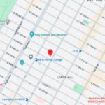
Upper East Side: Full Service Orthopedic Center & Walk-In
1198 3rd Avenue, Between 69th and 70th Street New York, NY 10021Physicians: Monday–Friday 8:00am–6:00pm, Saturday 8:00am–5:00pm
Physical Therapy: Monday–Friday 7:00am–7:30pm, Saturday 8:00am–3:00pm
Sunday: ClosedPhone: (212) 759-4553 -

Orthopedic Doctor Midtown Manhattan NYC — New York Bone & Joint Specialists
425 Madison Ave, Suite 200 (second floor) New York, NY 10017 (corner of East 49th Street)Physician appointments: Mon–Fri 8:00 am–6:00 pm
Physical therapy: Mon–Fri 7 am–7:30 pm, Sat 8 am–3 pmPhone: (212) 759-4553 -

Orthopedic Doctor Upper East Side NYC — New York Bone & Joint Specialists
130 E 67th St New York, NY 10065Currently ClosedPhone: (212) 759-4553