SLAP Repair & Shoulder Labrum Surgery NYC — Specialists at New York Bone & Joint
Written by: Dr. Popovitz.
Board-Certified Orthopedic Surgeon | Fellowship-Trained Sports Medicine, NYU Langone Medical Center | FAAOS
Co-Founder, NY Bone & Joint Specialists | Lenox Hill Hospital - Northwell Health
Recognized: New York Magazine Best Doctor | New York Times Super Doctor | IAOS Leading Physician of the World | IAOS Top Orthopedic Surgeon in New York
Date Published: May 5, 2026
Last Updated: June 3, 2026
Last Medically Reviewed: June 3, 2026
This page has been written and reviewed by a fellowship-trained, board-certified orthopedic surgeon with over 20 years of clinical experience. All clinical claims are supported by peer-reviewed literature and current AAOS and AOSSM guidelines. See References section below.
At a glance
- What it is: Arthroscopic surgery to repair a torn superior labrum — the cartilage ring at the top of the shoulder socket where the biceps tendon attaches. The repair reattaches the torn labrum using suture anchors, restoring the biceps anchor and eliminating the clicking, pain, and weakness that characterize a SLAP tear.
- What it treats: Type II SLAP tears (most common, highest surgical relevance), Type III and IV tears, labral tears adjacent to the biceps anchor, and paralabral cysts. Bankart lesions and anterior labral tears are a separate condition — see our dedicated Bankart repair page.
- Who performs it: Dr. Leon Popovitz and the New York Bone & Joint fellowship-trained surgical team, board-certified orthopedic surgeons with subspecialty training in shoulder surgery and 20+ years of SLAP repair and biceps tenodesis experience. Surgery at Lenox Hill Hospital.
- Where: Consultations, imaging, and physical therapy at two Manhattan offices: Upper East Side (1198 Third Ave) and Midtown (425 Madison Ave). Surgery at Lenox Hill Hospital.
- Recovery: Sling 4 weeks. Return to desk work: 2–3 weeks. Return to light overhead activity: 4–6 months. Return to competitive throwing or overhead sport: 6–9 months. All clearance is criteria-based, not time-based alone.
- New York Bone & Joint outcomes: Based on our own surgical outcome data, 90% of New York Bone & Joint overhead athlete patients return to competitive throwing, overhead sport or work after SLAP repair — compared to published return-to-sport rates of 40–80% for pitchers in the literature. [4] [5]
- Insurance: Most major insurance accepted. Coverage for SLAP repair is typically covered when medically indicated. Coverage verified before your procedure.
Book a consultation
Prompt Appointments Available
Upper East Side: 1198 Third Ave | Midtown: 425 Madison Ave
Surgery performed at Lenox Hill Hospital - Northwell Health
About this page
This page was written and is maintained by Leon Popovitz, MD, and the New York Bone & Joint surgical team. New York Bone & Joint Specialists is a private orthopedic surgery and sports medicine practice with two Manhattan locations: Upper East Side and Midtown. Dr. Popovitz is fellowship-trained in sports medicine at NYU Langone Medical Center and has specialized in shoulder surgery, including SLAP repair and biceps tenodesis, for over 20 years. SLAP repair and labrum surgery are performed by Dr. Popovitz and the New York Bone & Joint surgical team at Lenox Hill Hospital, part of the Northwell Health system. Dr. Popovitz served as former team physician for the US Open Tennis Championships and has treated overhead and throwing athletes at professional, collegiate, and recreational levels. New York Bone & Joint is independent of hospital systems and operates as a private practice serving patients across New York City and the surrounding region.
Introduction
A SLAP tear is among the most commonly misdiagnosed shoulder injuries in orthopedic medicine. The pain (deep in the shoulder, worse with overhead activity, sometimes accompanied by a click or catching sensation) overlaps with rotator cuff pathology, impingement, and biceps tendon problems. Standard MRI misses a significant percentage of SLAP tears. And the treatment decision, once the diagnosis is established, requires genuine subspecialty depth: the choice between repair and biceps tenodesis is one of the most nuanced in shoulder surgery, and getting it wrong has measurable consequences for recovery, reoperation risk, and return to sport. [1]
At New York Bone & Joint Specialists, our fellowship-trained shoulder surgeons have diagnosed and treated labral tears and SLAP pathology for over 20 years. Based on our own surgical outcome data, 90% of our overhead athlete patients return to competitive throwing or overhead sport after SLAP repair, compared to published return-to-sport rates of 40–80% for pitchers in the literature. [4] [5] That gap reflects our approach to diagnosis, our evidence-based decision-making between repair and tenodesis, and a rehabilitation protocol coordinated directly with our in-house physical therapy team.
If you have been told you have a SLAP tear or if you have had shoulder pain that no one has been able to explain, this page will give you a complete picture of what SLAP tears are, how they are accurately diagnosed, when surgery is the right answer, and exactly what the evidence says about your treatment options.
What is the Labrum: and What is a SLAP Tear?
The labrum is a ring of fibrocartilage that lines the rim of the shoulder socket (glenoid), deepening it and providing a stable surface for the humeral head to move against. Without the labrum, the shoulder socket would be too shallow to keep the ball of the humerus properly engaged during the extreme range of motion the shoulder demands. [1]
The biceps tendon (specifically the long head of the biceps) anchors to the top of the labrum at a structure called the biceps anchor. A SLAP tear (Superior Labrum Anterior to Posterior) is a tear of the labrum at this anchor point, at the very top of the shoulder socket, running from front to back. When the biceps anchor is torn or detached, the shoulder loses a key stabilizing structure, and the biceps tendon itself becomes a source of pain with every overhead movement.
The labrum is also continuous around the rest of the glenoid rim. Tears in the anterior (front) portion of the labrum (typically caused by shoulder dislocation) are called Bankart lesions and represent a distinct clinical entity with different surgical management. If you have been told you have a Bankart lesion or anterior labral tear, see our dedicated Bankart Repair & Shoulder Stabilization page for detailed information specific to your condition.
SLAP Tear Classification: Types I Through IV
Not all SLAP tears are the same. The Snyder classification system describes four types, each with different clinical significance and treatment implications.
| Type | Description | Clinical Significance | Treatment Direction |
| Type I | Fraying and degeneration of the superior labrum with an intact biceps anchor. Labrum is worn but not detached. | Often an incidental finding on MRI, particularly in older patients. May not be the source of symptoms. | Debridement (trimming of frayed tissue). Rarely requires formal repair. Often treated non-operatively. |
| Type II | Complete detachment of the superior labrum from the glenoid rim, the biceps anchor is destabilized. The most clinically significant and most common surgically treated SLAP tear. | This is the SLAP tear that causes symptoms in overhead athletes. The unstable biceps anchor produces pain, clicking, and weakness with overhead activity. | Surgical repair with suture anchors (SLAP repair) OR biceps tenodesis, depending on patient age, activity level, and intraoperative findings. See decision table below. |
| Type III | Bucket-handle tear of the superior labrum with an intact biceps anchor. The torn fragment displaces into the joint. | Produces mechanical symptoms: catching, locking. Biceps anchor remains stable. | Resection of the displaced bucket-handle fragment. Biceps anchor preserved. At New York Bone & Joint we make every effort to preserve and save your tissue: our goal is to repair the displaced labral tissue whenever tissue quality allows. |
| Type IV | Bucket-handle tear extending into the biceps tendon itself. Both labrum and tendon are involved. | Most severe. Biceps tendon integrity is compromised. Produces significant mechanical and functional symptoms. | Repair of the labrum and tendon, or biceps tenodesis depending on the extent of biceps tendon involvement and patient profile. |
“A key clinical nuance: Type II SLAP tears are the most surgically relevant, but they are also the most technically demanding to diagnose accurately. Standard MRI has limited sensitivity for SLAP tears, particularly for partial detachments and early Type II tears where the labrum has not fully separated from the glenoid. Very often we see patients come in with open MRI’s or “stand up” MRIs that are not powerful enough to detect the labra/SLAP. We routinely utilize high field MRIs that are either the newer 1.5T or, ideally, the 3T high field MRI to best visualize the labrum. Sometimes, we may have to use an MRI arthrogram (a contrast-enhanced study in which contrast is injected into the joint before imaging) to significantly improve diagnostic accuracy. [11] These are the steps that most frequently establishes the diagnosis that prior providers missed.” — Leon Popovitz, MD
Who Gets SLAP Tears and What Causes Them
SLAP tears affect two primary patient populations, through two distinct mechanisms:
- Overhead and throwing athletes: The most common population for symptomatic Type II SLAP tears. In throwers, the combination of extreme external rotation during the cocking phase and the forceful deceleration of the follow-through creates repetitive tensile load on the biceps anchor. Over time, this produces a peel-back mechanism. The biceps anchor progressively detaches from the glenoid with each throw. Baseball pitchers, tennis players, volleyball players, swimmers, and quarterbacks are particularly susceptible.
- Acute traumatic injury: A fall on an outstretched arm, a sudden traction injury to the arm, or a shoulder dislocation can produce an acute SLAP tear, often as part of a more complex labral injury. These patients are frequently younger and the tear is more discrete, making them better candidates for primary repair.
- Degenerative: In patients over 40, labral fraying at the biceps anchor is common and is often an MRI finding rather than the primary pain generator. Distinguishing a symptomatic Type II SLAP tear from an incidental degenerative finding in this population is one of the most challenging diagnostic tasks in shoulder medicine, and one where clinical examination and selective use of diagnostic injections plays a critical role.
Diagnosis: Why Standard MRI is Often Not Enough
Diagnosing a SLAP tear accurately requires more than ordering an MRI. The clinical examination (including provocation tests such as the O’Brien test, the biceps load test, and the compression-rotation test) establishes the index of suspicion. Imaging then confirms or characterizes the tear. The problem is that standard low field MRIs or open MRIs (the test most patients receive first) have limited sensitivity for SLAP tears, with studies showing they miss a clinically significant proportion of Type II tears. [11]
High field 3T closed MRI’s (and now some newer 1.5T MRs or even an MRI arthrogram, in which contrast is injected directly into the shoulder joint before imaging) significantly improves sensitivity and specificity for SLAP tears. The tear becomes visible on imaging that would otherwise appear normal. [11] At New York Bone & Joint, we recommend high field MRI or MRI arthrogram when: the clinical examination is strongly suggestive of a SLAP tear but standard low field or open MRI is negative or equivocal; the patient has had persistent shoulder symptoms without a clear structural diagnosis; or when we are evaluating for a labral tear in an overhead athlete with characteristic symptoms.
“The most common scenario I see is a patient who has had standard low field MRI or open MRI that reads as normal or showing minor bursitis or ‘mild degenerative changes.’ They have been told their shoulder looks fine on imaging and their pain is managed with cortisone injections or physical therapy that isn’t helping. I often have seen very young patients in their twenties and thirties come in with such MRI findings and treatment plans. That worries me because these patients are very young, and the repeated cortisone injections into their joint can cause degeneration of the cartilage. When I order the high field MRI or MRI arthrogram (with contrast inside the joint) the tear becomes visible. The diagnosis was there the whole time. The imaging was not the correct choice, and the treatment was not beneficial. Doctors take an oath to do no harm. Multiple cortisone injections into a young shoulder is not ideal.” — Leon Popovitz, MD
SLAP Repair vs. Biceps Tenodesis: The Most Important Decision
This is the decision that determines your outcome more than any other. Both procedures can successfully eliminate SLAP tear symptoms. The question is which one is right for your specific situation, and the answer depends on your age, your sport, and what the tissue looks like intraoperatively.
| What the evidence shows: Studies comparing SLAP repair to biceps tenodesis consistently show no significant difference in functional outcome scores (ASES, SANE, VAS pain scores) between the two procedures in appropriately selected patients. [6] [7] This means the choice isn’t about which procedure produces a better overall result but rather which procedure is the right fit for the individual patient profile. |
| Patient Profile | Primary Recommendation | Clinical Rationale & Evidence |
| Age < 30–35 | SLAP Repair | Recreates original anatomy. Traditionally preferred for younger, active patients with healthy labral tissue and an intact biceps anchor. Best outcomes in patients with acute traumatic detachment and good tissue quality. |
| Age > 35–40 | Biceps Tenodesis | SLAP repair in this age bracket is associated with significantly higher failure and reoperation rates in multiple studies, with some reporting up to a 1.45x higher failure risk compared to tenodesis. [7] [8] Tenodesis provides equivalent functional outcomes (ASES, SANE, VAS) with a lower reoperation rate and more predictable healing. |
| Overhead Athletes (age <30) | SLAP Repair (with careful counseling) | Repair is preferred to restore the native biceps anchor essential for throwing stability. However, return-to-sport rates in pitchers are variable in the literature: published rates of 40–80% for return to same level. [4] [5] New York Bone & Joint’s 90% return-to-overhead-sport outcome reflects careful patient selection and criteria-based rehabilitation. Patients must understand the recovery timeline. |
| Overhead Athletes (age >30) | Biceps Tenodesis | Tenodesis offers more reliable pain relief and faster return to sport in older throwers, particularly those with concurrent rotator cuff wear or degenerative labral changes. The trade-off of not restoring the native anatomy is well-tolerated in this population. |
| Degenerative SLAP (any age) | Biceps Tenodesis or Debridement | A Type I SLAP with fraying and degeneration but no true detachment typically does not require formal repair. For Type II tears with degenerative tissue, tenodesis (or debridement alone if the tissue quality is very poor) is preferred over attempting repair of tissue that will not hold suture anchors reliably. |
| Concurrent with rotator cuff repair | Debridement or Tenodesis | Performing SLAP repair concurrently with a full rotator cuff repair has been associated with increased post-operative stiffness. In patients undergoing rotator cuff repair who have SLAP pathology, debridement or tenodesis is generally preferred to a formal SLAP repair. |
“The repair vs. tenodesis decision may ultimately be made intraoperatively in many cases. The MRI tells us the tear is there and gives us a sense of the anatomy. But the tissue quality, such as how well the labrum holds suture, how robust the detachment is, and whether the tissue is healthy or degenerated, is assessed by feel and direct visualization once we are inside the joint. We go in with a plan based on your age, sport, and imaging, and we are prepared to adapt that plan based on what the tissue tells us. That intraoperative judgment is something that cannot be replicated by an algorithm or a protocol. Ultimately, we make every effort to recreate your original anatomy and preserve it. For this reason, I typically prefer repairing the labrum instead of a biceps tenodesis, which would mean cutting the biceps tendon and re-attaching in a location that is different than its original anatomy.” — Leon Popovitz, MD
Are You a Candidate for SLAP Surgery?
Not every SLAP finding on MRI requires surgery. The diagnosis must be confirmed, the symptoms must be attributable to the SLAP tear rather than concurrent pathology, and conservative management must have been appropriately tried. [1]
Surgery is typically recommended when:
- You have a confirmed Type II SLAP tear on MRI with symptoms consistent with the tear, such as pain with overhead activity, clicking or catching in the shoulder, biceps anchor tenderness, and a positive clinical examination.
- You are an overhead or throwing athlete whose symptoms prevent return to sport after an appropriate period of rest and physical therapy.
- You have failed a structured course of physical therapy and activity modification (typically 3–6 months).
- You have a Type III or IV SLAP tear with mechanical symptoms requiring surgical treatment of the displaced fragment.
- You have a labral tear with an associated paralabral cyst that is causing nerve compression or contributing to shoulder weakness. A cyst usually develops when the tear is large or long standing and the joint fluid travels through the tear.
Surgery is generally not recommended for Type I SLAP tears (fraying without detachment), incidental SLAP findings without correlating symptoms, or patients who have not completed an appropriate trial of conservative treatment. For older patients with degenerative Type II tears, the non-surgical pathway (physical therapy focused on posterior capsule stretching, rotator cuff strengthening, and scapular stabilization) should be thoroughly exhausted before any surgical discussion.
| From Dr. Popovitz: Fourteen Months to a Diagnosis A 29-year-old recreational tennis player came to see me after 14 months of right shoulder pain. I performed a labral/SLAP repair on his other shoulder about 5 years prior to that visit. He moved to another state after his surgery with me and went to see another orthopedic surgeon in his new city at a highly respected institution. He said that the new shoulder pain seemed very similar to the shoulder pain as he had on his left shoulder, which I fixed. The new, highly respected orthopedic surgeon diagnosed him with a Type II SLAP tear and impingement (bone spur causing bursitis). The patient then had physical therapy for 2 months which did not help. So, he underwent a right shoulder arthroscopy with an acromioplasty/decompression (removal of the bone spur) and debridement of the SLAP tear. The pain never went away, and he decided to travel back to New York to see me. When I saw the patient, he told me that he was concerned because he had a new baby and he had difficulty lifting his child. I examined his right shoulder. The O’Brien test (a test on physical exam) was strongly positive, and his biceps load test was positive. I ordered a new MRI arthrogram to best visualize how much tissue was remaining after the debridement and if the tear was present (as I suspected). The MRI arthrogram results showed the Type II SLAP tear, the development of a paralabral cyst (possibly from the long standing tear) and evidence of the performed acromioplasty (removal of a portion of bone under the acromion). Typically, a bone spur isn’t common in such a young patient, and we don’t routinely rush to do an acromioplasty in a young patient. Moreover, a Type II SLAP tear that has failed conservative treatment is usually repaired for young active patients instead of only debrided. Again, the goal is to preserve the tissue. I performed an arthroscopic SLAP/labral repair with a cyst drainage. The patient was 29, he had healthy tissue, and an acute-pattern tear: a textbook candidate for repair. At eight months post-surgery, he was back to playing recreational tennis with no limitations. What stays with me from this case is the fourteen months that the man had to suffer and the difficulty he had even raising and enjoying his newborn child. I am so grateful I was able to relieve his suffering, allow him to return to his life and to preserve his tissue for a long lifetime to come. — Leon Popovitz, MD |
The Procedure: What Happens During SLAP Repair
SLAP repair with New York Bone & Joint is performed arthroscopically under general anesthesia, typically combined with an interscalene nerve block for post-operative pain control and to limit the amount of general anesthesia. Most procedures take 45–90 minutes. Patients go home the same day. [3]
Your surgeon will:
- Create two or three small portals around the shoulder, typically less than a centimeter each.
- Perform a complete arthroscopic survey of the entire shoulder: the labrum, the biceps anchor, the rotator cuff, the subacromial space, the articular surfaces, and the joint capsule. Concurrent pathology is identified and documented.
- Assess the tear and the tissue: The labrum is probed to assess the degree of detachment, the quality and pliability of the tissue, and the vascularity of the tear. This intraoperative assessment helps determine whether repair or tenodesis proceeds, and for a Type II tear, whether the tissue is robust enough to hold suture anchors reliably.
- For SLAP repair: The glenoid rim at the biceps anchor is prepared to create a vascular bed. Suture anchors are placed in precise positions at the superior glenoid, and sutures are passed through the labrum to reattach it to bone. The number of anchors depends on the extent of the tear,
- For biceps tenodesis: The biceps tendon is released from its attachment at the superior labrum and reattached to the proximal humerus below the shoulder joint, eliminating the tension on the damaged labral anchor. This can be performed arthroscopically or through a small open incision depending on the preferred fixation technique.
- Address any concurrent findings (such as rotator cuff pathology, Bankart component, loose bodies, or subacromial impingement) in the same operative session.
- Close the portals and apply a sling. You leave with clear post-operative instructions and a physical therapy protocol.
“One nuance that shapes every surgery that I perform: I always focus deeply on saving the patient’s own tissue. My preference is to repair your labrum so that you can live with your original anatomy. I recognize the role and purpose of tenodesis and in some circumstances it is absolutely necessary, such as when the biceps itself is severely damaged. Nevertheless, whenever possible my preference is to repair your labrum and allow your biceps to stabilize naturally as a result of the repair instead of cutting the biceps and reattaching it in a different anatomical position.” — Leon Popovitz, MD
| SLAP Tears vs. Bankart Lesions: Related but Distinct Both SLAP tears and Bankart lesions are labral injuries, but they affect different parts of the labrum, arise from different mechanisms, and require different surgical approaches. SLAP tear: Superior labrum tear at the biceps anchor. Caused by overhead mechanics, traction injury, or fall on outstretched arm. Produces pain and clicking with overhead activity. Treated with SLAP repair or biceps tenodesis depending on patient profile. Bankart lesion: anterior labral tear at the 3–6 o’clock position, caused by anterior shoulder dislocation. Produces instability, apprehension, and recurrent subluxation or dislocation. Treated with arthroscopic Bankart repair to restore the shoulder’s anterior restraint mechanism. The two can occur together. A shoulder dislocation can cause both a Bankart lesion and a superior labral component. When both are present, they are addressed in the same procedure. |
Recovery and Rehabilitation
Recovery after SLAP repair is slower than most patients expect, and for good reason. The repaired labrum must undergo a biological healing process before it can tolerate load. The rehabilitation protocol is carefully staged to protect the repair during healing while preventing the shoulder stiffness that is the primary complication of over-immobilization.
| Phase | Timeframe | Goals & Activities |
| Protection | Weeks 0–4 | Sling worn continuously. Pendulum exercises begin early to prevent stiffness. No active use of the repaired arm. Return to desk work typically by week 2–3. This phase is critical: placing load on the biceps anchor before the repair has healed is the primary mechanism of early failure. |
| Passive Motion | Weeks 4–8 | Progressive passive range-of-motion under PT direction. Gradual elevation and rotation. Sling weaned as directed by your surgeon. No active biceps use yet. |
| Active Motion | Weeks 8–12 | Active range-of-motion begins. Strengthening of the periscapular muscles and rotator cuff. No biceps-loading exercises (curls, heavy gripping) until specifically cleared. |
| Strengthening | Months 3–6 | Progressive shoulder strengthening. Sport-specific conditioning begins. For throwing athletes, an interval throwing program starts under physical therapist supervision. |
| Return to Sport | Months 6–9 | Return to overhead sport and competitive throwing with criteria-based clearance: strength symmetry testing, range-of-motion documentation, and physician sign-off. Not time-based alone. [9] New York Bone & Joint’s 90% return-to-throwing/work outcome is built on this structured, criteria-based progression. |
Biceps tenodesis recovery follows a similar but slightly faster trajectory in many patients, particularly for the return-to-activity phase. Because the biceps tendon is reattached at a lower-tension site, the protection requirements are less strict and progression can occur slightly earlier. Your surgeon will provide a specific protocol based on the procedure performed.
In-House Physical Therapy: Why it Matters Especially After SLAP Surgery
SLAP repair has a narrower rehabilitation window than most shoulder procedures. Too aggressive early and you risk re-tearing the repair. Too conservative and you develop the shoulder stiffness that is the most common post-operative complication of over-immobilization. The protocol must be calibrated to what was done in the operating room: which anchors were placed, where the tear was, how robust the repair was, and whether a tenodesis was performed.
At New York Bone & Joint, your physical therapist and surgeon work in the same center and communicate directly from day one. Before your first physical therapy session, your therapist has reviewed your operative report and confirmed the specific repair type and protocol with your surgeon. When questions arise during your recovery, they are answered the same day.
For throwing and overhead athletes, our return-to-sport progression follows a sport-specific interval program with objective functional testing at each milestone. Clearance to return to competitive throwing requires passing strength symmetry assessments and a staged interval throwing program, not just reaching a time threshold. This structured progression is a core component of our 90% return-to-throwing/work outcome.
Risks and Considerations
SLAP repair is a safe procedure in experienced hands. The overall complication rate for shoulder arthroscopy is less than 1%. [10] Specific risks to understand
- Re-tear or repair failure: The primary risk specific to SLAP repair. In younger patients with acute tears and good tissue quality, failure rates are very low. In patients over 35–40 where the evidence supports tenodesis as having lower repair failure and reoperation rates, the decision between procedures is consequential. [7] [8] But beyond age and tissue quality, outcomes are directly shaped by the surgeon’s experience and success with both techniques. In the right hands, either procedure can produce excellent results. This is why the repair vs. tenodesis decision, and the experience of the surgeon performing it, is the most important part of your pre-operative consultation.
- Stiffness: The most common complication of SLAP repair. Over-immobilization during the protection phase leads to adhesive capsulitis-like changes. Early pendulum exercises and a carefully staged passive motion protocol significantly reduce this risk. Your physical therapy protocol is designed specifically to prevent stiffness while protecting the repair.
- Incomplete return to overhead sport: Particularly for high-level pitchers. Published return-to-sport rates for professional pitchers range from 40–80%, and return to the same level of performance is even more variable. [4] [5] New York Bone & Joint’s 90% return-to-overhead-sport outcome/work reflects our successful experience, patient selection, repair/tenodesis decision framework, and rehabilitation protocol. Honest pre-operative counseling about realistic expectations is part of every consultation.
- Infection: Rare. Less than 1% with standard arthroscopic technique and sterile protocol. [10]
- Nerve injury: Very rare with standard arthroscopic portals for SLAP repair. Discussed in detail for complex cases.
At your consultation, your surgeon will walk through the specific risks relevant to your tear type, your age, your activity level, and your individual health profile.
Why Choose New York Bone & Joint for SLAP Repair?
| New York Bone & Joint Advantage | What It Means for You |
| 90% return-to-overhead-sport/work (vs. published 40–80%) | Based on our own surgical outcome data. Built on evidence-based repair vs. tenodesis decision-making, careful patient selection, and a structured criteria-based rehabilitation protocol. |
| Evidence-based SLAP vs. tenodesis framework | We use published evidence: ASES, SANE, VAS equivalence; age-stratified failure risk data: to make the right procedure recommendation for your profile. We select the procedure best matched to your age, tissue quality, and activity profile, drawing on our experience and success with both techniques. |
| High field MRI or MRI arthrogram as standard for equivocal cases | Open or low field MRIs miss a clinically significant percentage of SLAP tears. When the examination is positive and the open or low field MRI is negative, we order the high field MRI or arthrogram. This is the step that most often establishes a diagnosis that prior providers missed. [11] |
| Intraoperative tissue judgment | The repair vs. tenodesis decision is finalized intraoperatively based on tissue quality. A plan made in the office must be adaptable to what the tissue shows inside the joint. |
| In-house PT — same center | Your surgeon and physical therapist communicate directly. The protocol is calibrated to your specific repair. The stiffness prevention and return-to-throwing progression are coordinated from day one. |
| Honest return-to-sport counseling | We tell overhead athletes the truth about recovery timelines and realistic outcomes before surgery, not after. For high-level pitchers, that conversation includes the published data and our own outcomes. |
| Same surgeon, start to finish | The physician who evaluates you is the surgeon who operates on you. No handoffs. |
References
- American Academy of Orthopaedic Surgeons. SLAP Tears. OrthoInfo. orthoinfo.aaos.org/en/diseases–conditions/slap-tears
- American Academy of Orthopaedic Surgeons. Biceps Tendon Tear at the Shoulder. OrthoInfo. orthoinfo.aaos.org/en/diseases–conditions/biceps-tendon-tear-at-the-shoulder
- American Academy of Orthopaedic Surgeons. Shoulder Arthroscopy. OrthoInfo. orthoinfo.aaos.org/en/treatment/shoulder-arthroscopy
- Thayaparan A et al. Return to sport following arthroscopic SLAP repair in overhead athletes. Sports Health. 2019. pubmed.ncbi.nlm.nih.gov/31584340
- Fedoriw W et al. Return to play after treatment of superior labral tears in professional baseball players. Am J Sports Med. 2014. pubmed.ncbi.nlm.nih.gov/24674945
- Song Y et al. Arthroscopic Modified Double-Row Biceps Tenodesis versus Labral Repair for the Treatment of Isolated Type II SLAP Lesions in Non-Overhead Athletes. Orthop Surg. 2022. pubmed.ncbi.nlm.nih.gov/35633041
- Provencher MT et al. A prospective analysis of 179 type 2 superior labrum anterior and posterior repairs: outcomes and factors associated with success and failure. Am J Sports Med. 2013. pubmed.ncbi.nlm.nih.gov/23460326
- Neri BR et al. Outcome of type II superior labral anterior posterior repairs in elite overhead athletes: Effect of concomitant partial-thickness rotator cuff tears. Am J Sports Med. 2011. pubmed.ncbi.nlm.nih.gov/20940452
- Elsenbeck MJ et al. Return to Sports After Shoulder Stabilization Surgery for Anterior Shoulder Instability. Curr Rev Musculoskelet Med. 2017. pubmed.ncbi.nlm.nih.gov/29076041
- Shin JJ et al. Complications after arthroscopic shoulder surgery. J Am Acad Orthop Surg Glob Res Rev. 2018. pubmed.ncbi.nlm.nih.gov/30680371
- Momenzadeh OR et al. Assessment of Correlation Between MRI and Arthroscopic Pathologic Findings in the Shoulder Joint. Arch Bone Jt Surg. 2015. pmc.ncbi.nlm.nih.gov/articles/PMC4628637
FAQs
A SLAP tear is a tear of the superior labrum (the cartilage at the top of the shoulder socket) at the point where the biceps tendon attaches, called the biceps anchor. SLAP stands for Superior Labrum Anterior to Posterior, describing the location of the tear. The most clinically significant type is a Type II SLAP tear, in which the biceps anchor is fully detached from the glenoid rim, producing pain, clicking, and weakness with overhead activity. [1]
The classic symptoms of a SLAP tear are deep shoulder pain that worsens with overhead activity, a clicking or catching sensation in the shoulder, and weakness or instability with throwing or reaching overhead. A positive O’Brien test or biceps load test on clinical examination raises the suspicion significantly. Standard open or low field MRIs can miss SLAP tears. High field MRI or MRI arthrogram are significantly more sensitive and specific and is recommended when clinical examination is positive but standard MRI is negative or equivocal. [11]
The answer depends primarily on your age and the surgeon’s experience: for patients under 30–35 with acute tears and healthy tissue, SLAP repair is generally preferred; for patients over 35–40, biceps tenodesis typically provides equivalent functional outcomes with a significantly lower reoperation rate. Studies comparing the two procedures show no significant difference in ASES, SANE, or VAS pain scores in appropriately selected patients. [6] [7] The decision is also made intraoperatively. Tissue quality is assessed directly and the plan is adapted if the labral tissue will not support a durable repair. Ultimately, it also depends upon the surgeon’s experience and expertise.
Based on New York Bone & Joint’s own surgical outcome data, 90% of our patients return to throwing, overhead sport or work after SLAP repair, compared to published return-to-sport rates of 40–80% for pitchers in the literature. [4] [5] Return-to-sport for high-level pitchers is particularly variable in published studies, and honest pre-operative counseling about realistic expectations is part of every consultation at New York Bone & Joint. Our outcome reflects our experience, expertise, patient selection, the repair vs. tenodesis decision framework, and a structured, criteria-based rehabilitation protocol.
Most patients wear a sling for 4 weeks, return to desk work within 2–3 weeks, and return to overhead sport with criteria-based clearance at 6–9 months. Return to competitive throwing requires completing a staged interval throwing program and passing strength symmetry testing, not just reaching a time threshold. [9] Biceps tenodesis recovery follows a similar but slightly faster trajectory for daily activities.
A SLAP tear affects the superior (top) labrum at the biceps anchor; a Bankart lesion affects the anterior (front) labrum and is caused by shoulder dislocation. Both are labral injuries, but they arise from different mechanisms, produce different symptoms, and require different surgical approaches. SLAP tears cause pain and clicking with overhead activity. Bankart lesions cause shoulder instability, apprehension, and recurrent dislocations. The two can occur together, in which case both are addressed in a single procedure.
Standard open or low field MRI misses a clinically significant percentage of SLAP tears: MRI arthrogram is significantly more sensitive and is recommended when clinical examination is positive but standard MRI is negative or equivocal. [11] MRI arthrogram involves injecting contrast directly into the shoulder joint before imaging, which fills the space between a partially detached labrum and the glenoid, making the tear visible. At NYBJ, we order high field MRIs and an MRI arthrogram when the clinical picture warrants it and standard MRI is insufficient. We coordinate prompt scheduling at affiliated imaging facilities.
Type I SLAP tears (fraying without detachment) often do not require surgery and can be managed with physical therapy focused on rotator cuff and scapular strengthening. True Type II SLAP tears with detachment of the biceps anchor do not heal on their own, because the detached labrum loses its vascular supply. For symptomatic Type II tears, surgical treatment is typically required to restore function. However, not every Type II SLAP finding on MRI is symptomatic, and the decision to operate is based on the complete clinical picture, not the imaging finding alone.
Arthroscopic SLAP repair for confirmed Type II tears with documented symptoms and failed conservative treatment is typically covered by major insurance plans when medically indicated. Our team will verify your coverage before your procedure and provide a clear picture of your out-of-pocket responsibility, if any, in advance.
Medically Reviewed by Dr. Popovitz.
You can book your appointment with New York Bone & Joint online by submitting the appointment request form here:
Book an appointmentOur Locations
-
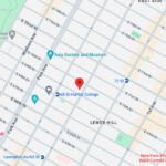
Upper East Side: Full Service Orthopedic Center & Walk-In
1198 3rd Avenue, Between 69th and 70th Street New York, NY 10021Physicians: Monday–Friday 8:00am–6:00pm, Saturday 8:00am–5:00pm
Physical Therapy: Monday–Friday 7:00am–7:30pm, Saturday 8:00am–3:00pm
Sunday: ClosedPhone: (212) 759-4553 -

Orthopedic Doctor Midtown Manhattan NYC — New York Bone & Joint Specialists
425 Madison Ave, Suite 200 (second floor) New York, NY 10017 (corner of East 49th Street)Physician appointments: Mon–Fri 8:00 am–6:00 pm
Physical therapy: Mon–Fri 7 am–7:30 pm, Sat 8 am–3 pmPhone: (212) 759-4553 -

Orthopedic Doctor Upper East Side NYC — New York Bone & Joint Specialists
130 E 67th St New York, NY 10065Currently ClosedPhone: (212) 759-4553